
Rapid culture-free diagnosis of clinical pathogens via integrated microfluidic-Raman micro-spectroscopy
Yuetao Li†; Jiabao Xu†; Xiaofei Yi; Xiaobo Li; Yanjun Luo; Andrew Glidle; Phil Summersgill; Simon Allen; Tim Ryan; Xiaochen Liu; Wei Yu; Xiaobing Chu; Shiyu Chen; Qian Zhang; Xiaogang Xu; Xiaoting Hua; Qiwen Yang; Julien Reboud; Yunsong Yu*; Wei E. Huang*; Jonathan M. Cooper*; Huabing Yin*
† Equal contribution* Corresponding authors
A platform combining microfluidics, Raman spectroscopy and deep learning identifies pathogens directly from clinical samples within 20 minutes. A 305-patient study found high agreement with culture methods; broader validation is needed before clinical implementation.
Key points
- Faster workflow. The integrated platform reported sample-to-report testing within 20 minutes.
- Chemical fingerprints. Single-cell Raman measurements and deep learning support pathogen identification.
- Clinical evaluation. A 305-patient study found 95.4% agreement with culture; broader validation remains necessary.
Preparing a sample without growing the pathogen
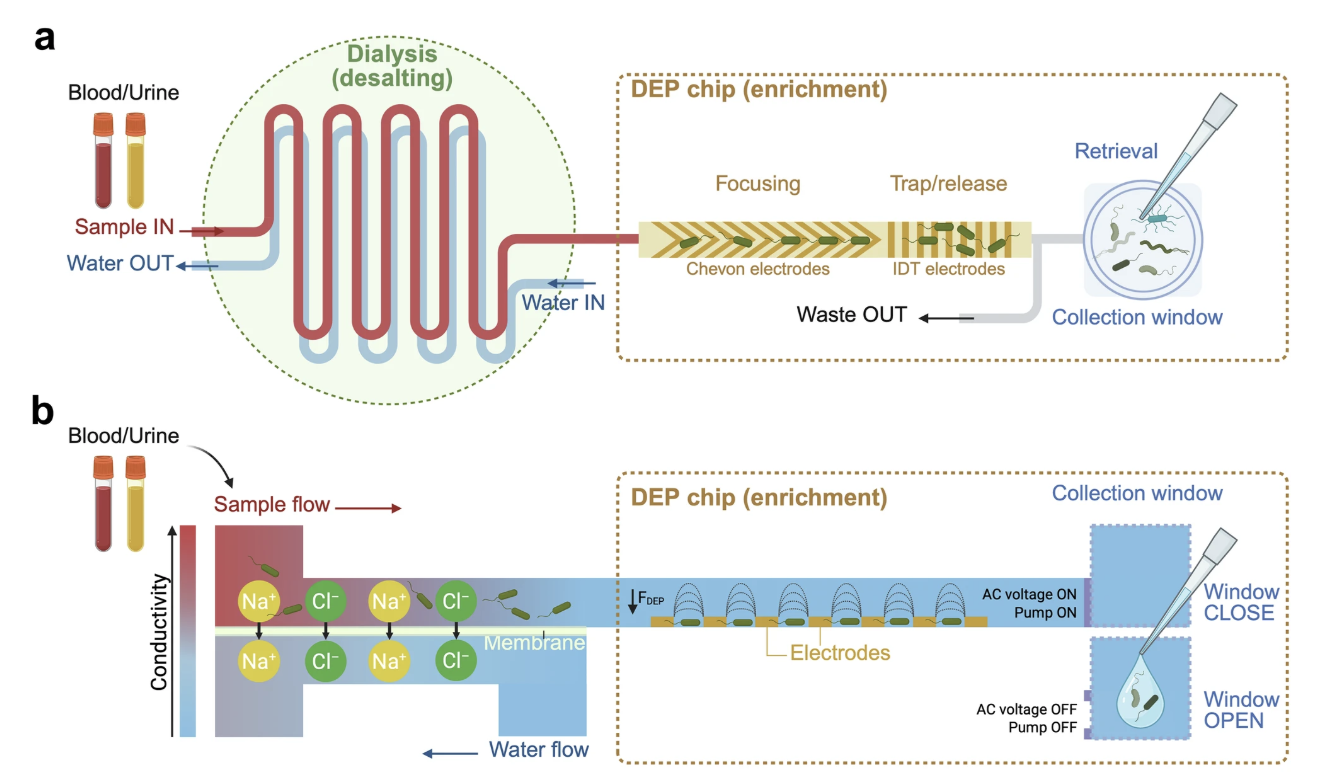
The platform addresses a practical bottleneck: clinical samples can contain too few microbial cells, mixed with material that interferes with measurement. Its first task is to isolate and concentrate those cells without waiting for a culture to grow. The researchers combined dialysis with dielectrophoresis, a method that uses a nonuniform electric field to capture particles such as microbial cells.
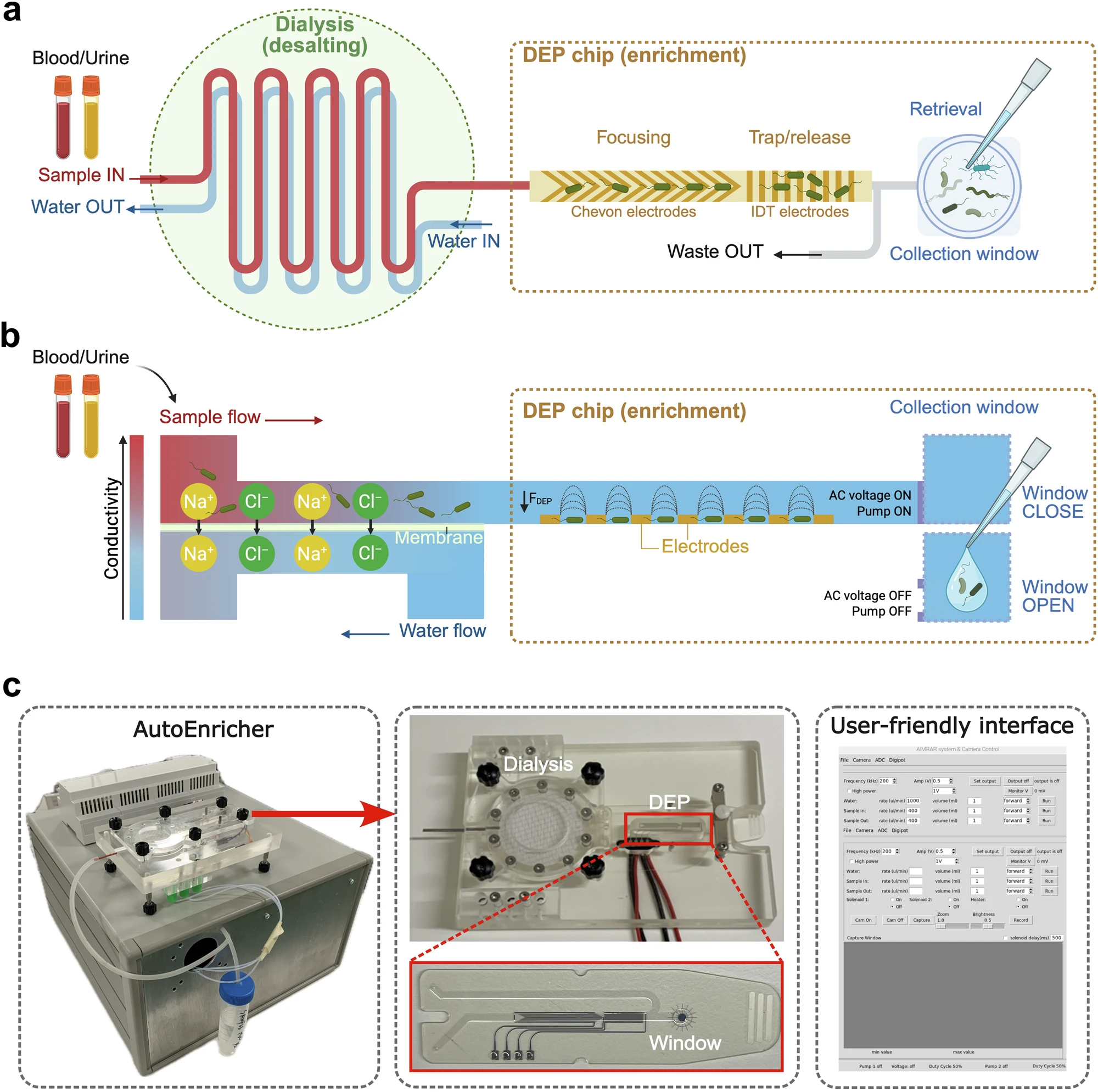
As the sample passes through the dialysis unit, salts diffuse across a membrane into a separate water stream. Lowering conductivity makes electrical capture more effective. The sample then enters a chip where electrodes collect pathogens; switching off the field releases them into a small collection window. Tests across additional bacterial and fungal species found capture efficiencies ranging from 89.1% to 99.5%.
 View full size
Figure 2. Dialysis reduces salt content before electrical capture concentrates microbial cells. The photographs below show the instrument and disposable components used to prepare samples for Raman analysis.
Li et al. (2025) · Original paper ↗ · CC BY 4.0 · Schematic created in BioRender by J. Xu.
Where the 20 minutes go
In the clinical workflow, approximately 1 mL of sample was concentrated into a roughly 3 microlitre droplet. Automated priming, enrichment and release took less than ten minutes in the reported workflow. A small portion was transferred to a slide for inspection, allowing the operator to assess whether microbial cells were present at the relevant threshold.
Samples classified as negative could finish at this stage, at approximately twelve minutes. Positive samples continued to Raman measurement and automated analysis. Recording spectra from around fifteen cells took about five minutes, with computational classification taking less than a minute. The reported turnaround therefore describes a sequence of sample preparation, triage and identification steps, rather than the speed of the AI model alone.
Turning single-cell spectra into an identification
Raman spectroscopy records molecular vibrations, producing a chemical fingerprint from each measured cell. The reference database contained 342 clinical isolates representing 29 bacterial and seven fungal species. A one-dimensional ResNet model learned to distinguish the spectral patterns, achieving 95.1% species-level accuracy in the laboratory isolate evaluation.
To adapt the model to clinical material, the researchers used spectra from forty confirmed single-infection urine samples for fine-tuning. Predictions from multiple cells were then combined at sample level. Measuring individual cells also allows different organisms within a mixed sample to be considered separately, rather than averaging all of their signals into one spectrum.
Two clinical results that answer different questions
The 305-patient evaluation at two hospitals first assessed positive or negative infection status against conventional culture. It reported 95.4% agreement, 98.5% sensitivity and 83.6% specificity. These numbers concern infection detection. They should not be read as the accuracy of identifying the exact pathogen species in all 305 samples.
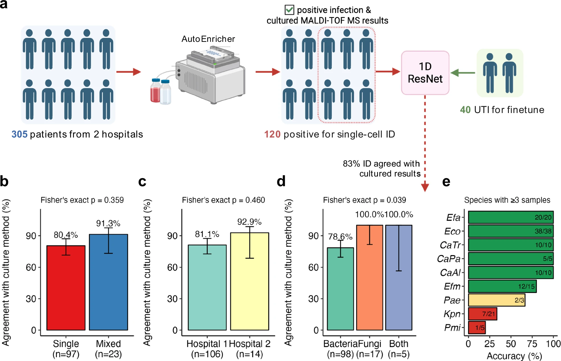
Species identification was assessed separately in 120 positive samples containing pathogens represented in the Raman database. This group included 97 single infections and 23 mixed infections. Investigators performing Raman measurements and classification were blinded to the reference results. Raman identification agreed with culture and MALDI-TOF mass spectrometry in 99 of the 120 samples: 82.5% overall agreement. Reporting these two stages separately shows where performance is already strong and where further improvement is needed.
 View full size
Figure 8. Species identification was assessed in 120 positive clinical samples, with 82.5% overall agreement against the reference method. This is separate from the 305-patient evaluation of positive or negative infection status.
Li et al. (2025) · Original paper ↗ · CC BY 4.0 · Schematic created in BioRender by J. Xu.
What the evaluation supports
The study demonstrates a route from an unprocessed clinical sample to rapid detection and, for positive samples, a proposed pathogen identity. Its strongest practical contribution is the integration: enrichment supplies measurable cells, spectroscopy captures their chemistry, and a trained model interprets those measurements. The spectra database, code and trained models were also made available through the publication’s data repository.
The clinical cohort was predominantly urine samples—298 of 305—so performance across other sample types needs broader evaluation. Fifteen additional blood-culture samples demonstrated technical feasibility with a complex matrix but were excluded from the culture-free cohort. Study results were not used to manage patients. Larger clinical studies, assessment of discordant results and integration with antimicrobial susceptibility testing are needed to establish how the workflow could support treatment decisions.
Publication
Rapid culture-free diagnosis of clinical pathogens via integrated microfluidic-Raman micro-spectroscopy
Yuetao Li†; Jiabao Xu†; Xiaofei Yi; Xiaobo Li; Yanjun Luo; Andrew Glidle; Phil Summersgill; Simon Allen; Tim Ryan; Xiaochen Liu; Wei Yu; Xiaobing Chu; Shiyu Chen; Qian Zhang; Xiaogang Xu; Xiaoting Hua; Qiwen Yang; Julien Reboud; Yunsong Yu*; Wei E. Huang*; Jonathan M. Cooper*; Huabing Yin*
† Equal contribution* Corresponding authors
Nature Communications · 2025
Read the original publication