
AI-enabled rhodopsin design for blue-light enhanced bacterial growth
Haris Saeed; Michael J. Lewis; Takayoshi Fujiwara; Jek Huang; Masae Konno; Kaho Mori; Susumu Yoshizawa; Keiichi Inoue; Tiezheng Pan; Yun Wang; Aidong Yang; Wei E. Huang*
* Corresponding author
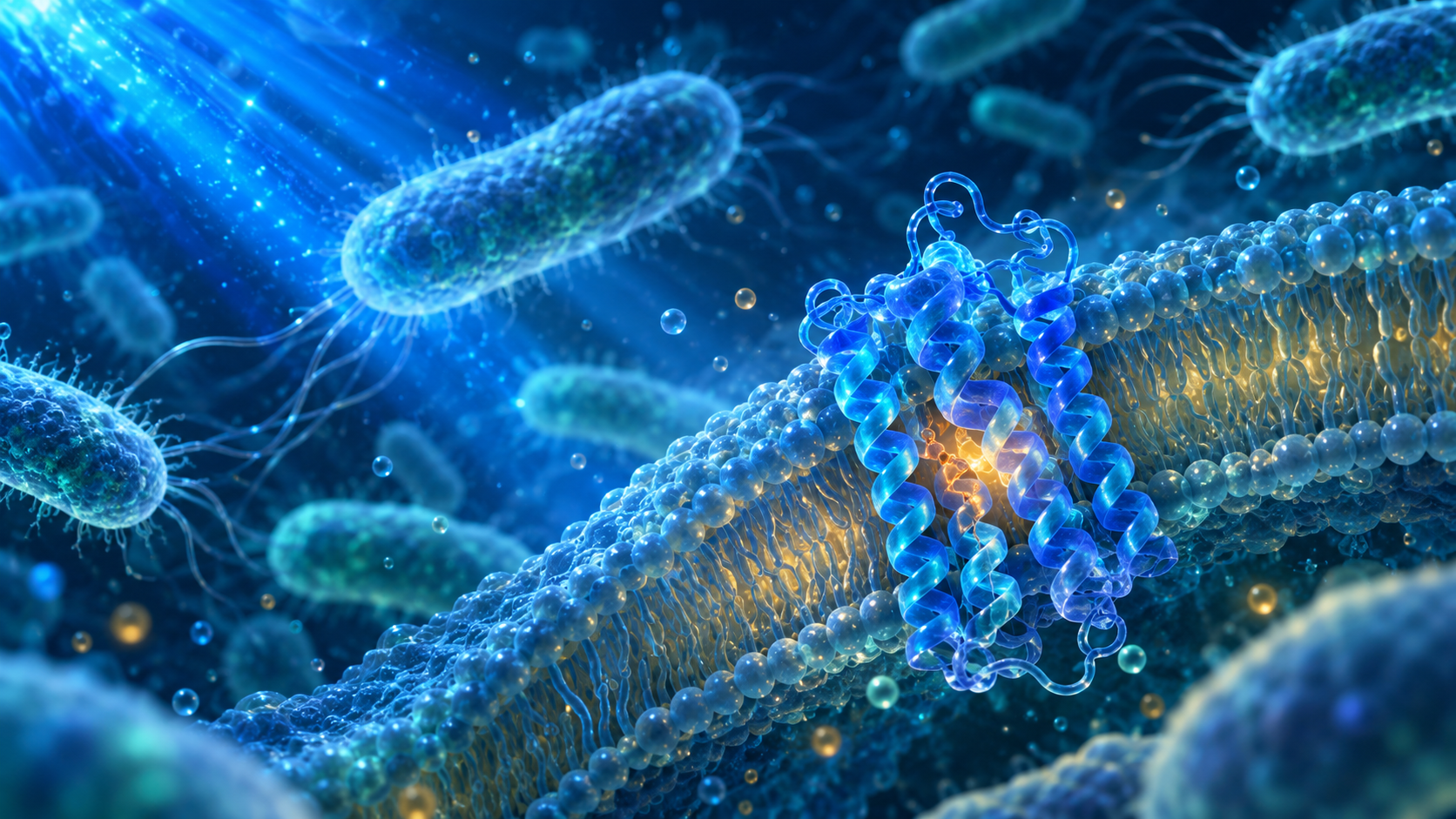
An AI-guided pipeline designed four rhodopsin variants that absorb blue light. Experiments showed enhanced growth of Cupriavidus necator under blue-light illumination. These findings are reported in a preprint and have not yet been peer reviewed.
Key points
- AI-guided design. A computational workflow generated four rhodopsin candidates for experimental testing.
- Blue-light response. All four variants absorbed light near 410 nm and enhanced bacterial growth under blue light.
- Research stage. The findings are reported in a preprint and have not yet been peer reviewed.
Searching beyond familiar proteins
Microbial rhodopsins contain retinal, a light-absorbing molecule held inside a protein pocket. Changes around that pocket can alter the wavelengths the protein absorbs. The challenge in this study was to explore a less familiar part of the spectrum while preserving sequence features associated with proton-pumping rhodopsins.
The researchers assembled a dataset of 884 rhodopsin sequences with measured absorption wavelengths. Of the 310 proton pumps in that collection, only 11 absorbed most strongly below 500 nm. Blue-shifted designs therefore lay in a sparsely sampled region, where predictions are harder to make. The computational goal was to find promising candidates for testing, rather than assume that a predicted wavelength would be exact.
How the AI workflow chooses candidates
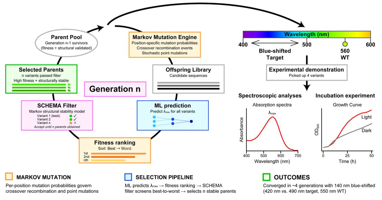
A genetic algorithm repeatedly proposes changes to protein sequences. A predictor combining LASSO and XGBoost estimates the wavelength each candidate might absorb, while a Markov model checks whether the sequence still resembles a proton pump. Candidates that fail this plausibility check cannot seed the next generation. This keeps the search from favouring a spectral prediction at the expense of the intended protein family.
For the experimental series, the search targeted 490 nm. Seven candidates with substantial sequence differences from the starting scaffold were selected across ten computational runs, then ranked using AlphaFold 3 structural predictions. Five were taken forward for cloning; four could be successfully expressed and became APR1, APR2, APR6 and APR7. The fifth did not yield viable transformants and was not characterised further.
 View full size
Figure 1. The design loop proposes protein sequences, predicts their absorption wavelengths and filters candidates before laboratory testing. The curves on the right illustrate the validation workflow.
What the absorption measurements revealed
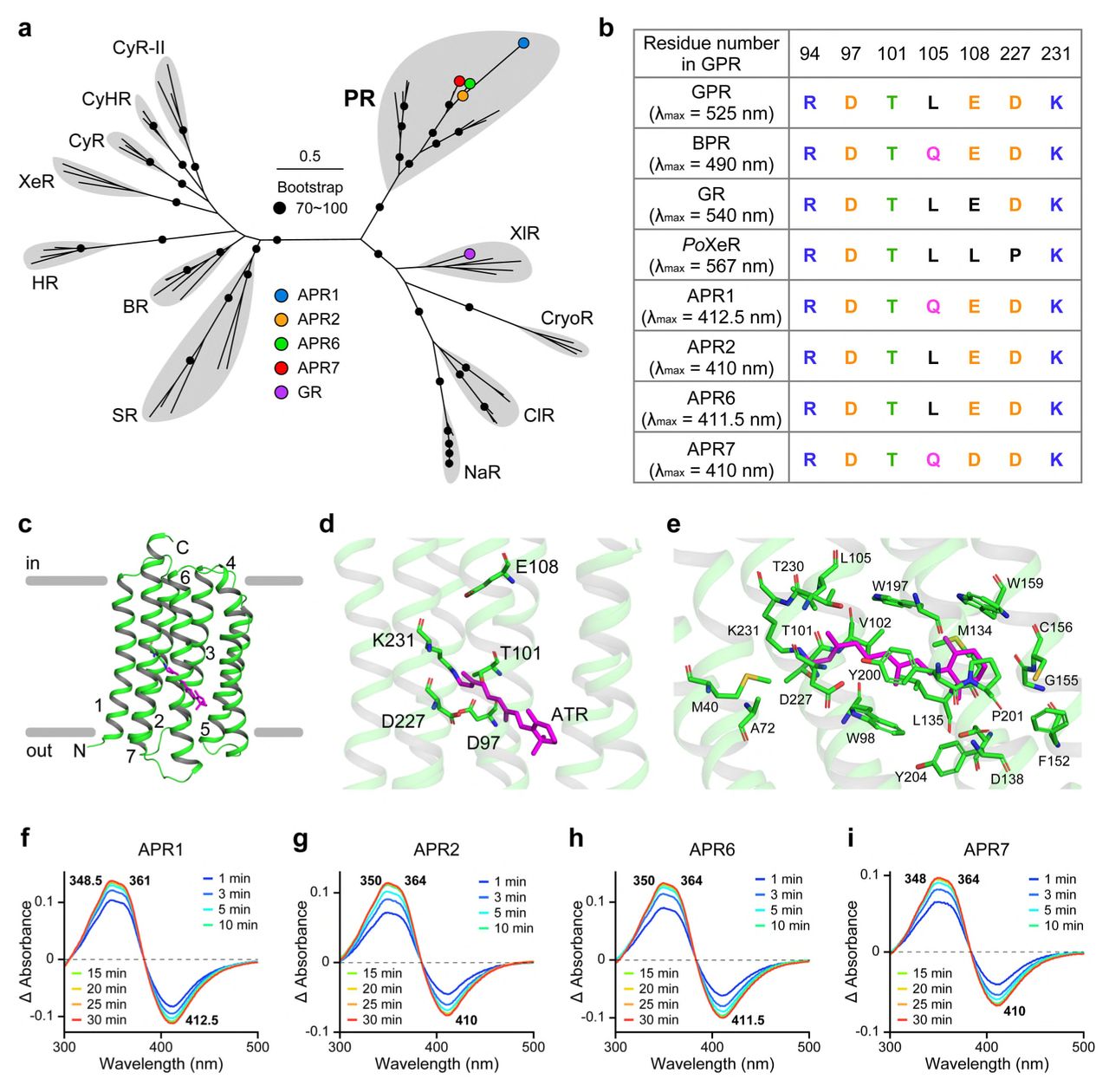
The team measured membrane fractions from E. coli expressing each design. They compared absorption before and after hydroxylamine treatment, which breaks the linkage between retinal and the protein. The resulting spectral change helps separate retinal-associated absorption from other components in the membrane preparation.
The measured peaks were 412.5 nm for APR1, 410 nm for APR2, 411.5 nm for APR6 and 410 nm for APR7—substantially shorter wavelengths than the 490 nm design target. This was a useful blue shift, but also showed the limits of precise wavelength prediction. AlphaFold models supported a rhodopsin-like architecture; the spectroscopy supplied experimental evidence that the designs incorporated retinal and absorbed near the violet end of the visible spectrum.
 View full size
Figure 5. Predicted structures and measured spectra of the four APR variants. The lower panels show absorption changes near 410 nm; these measurements support retinal binding but do not directly measure proton pumping.
Following growth under light and dark
The four proteins were also expressed in Cupriavidus necator H16. Growth was monitored for 50 hours in controlled bioreactors, comparing illuminated cultures with dark controls. The APR cultures received 457 nm blue light. The study also included a green-absorbing rhodopsin reference, illuminated separately with green light, and uninduced controls to help assess the effect of protein expression.
APR-expressing strains showed a sustained growth advantage under illumination. For example, APR1 reached a growth rate of 0.139 per hour at the 15-hour measurement under light, compared with 0.085 per hour in the dark. Across the exponential-growth window, all four APR strains grew faster than the pooled dark-induced control. These comparisons connect the protein designs to a measurable response in a living host.
What is established, and what comes next
Measurements of ATP and ADP also indicated a more energy-rich cellular state under illumination across the strains tested. However, the size of that light response did not differ significantly between strains. The energy measurements therefore support an association with illumination, rather than establishing that one APR design has a uniquely superior energy-conversion mechanism.
The work joins computational design, optical measurements and bacterial growth experiments in one study. Direct measurements of proton transport and photocycle behaviour remain important next steps: the bleaching assay does not measure pumping efficiency. The results provide a basis for developing light-responsive cell systems, while manufacturing productivity and performance at scale remain to be tested. This work is a preprint and has not yet been peer reviewed.
Publication
AI-enabled rhodopsin design for blue-light enhanced bacterial growth
Haris Saeed; Michael J. Lewis; Takayoshi Fujiwara; Jek Huang; Masae Konno; Kaho Mori; Susumu Yoshizawa; Keiichi Inoue; Tiezheng Pan; Yun Wang; Aidong Yang; Wei E. Huang*
* Corresponding author
bioRxiv · 2026 · Preprint, not peer reviewed
Read the original publication