
Raman Micro-spectroscopy
Deciphering metabolic logic through vibration: non-invasive diagnostics at the resolution of a single cell.
Diagnostic window
Our lab pioneered the use of stable-isotope probing paired with Raman to map functional metabolic active cells within complex microbiome communities.
Phenotype to genotype
Finding cells by their chemical fingerprint
A single-cell Raman spectrum records molecular vibrations from one cell. Its pattern can reveal biochemical features without first attaching a fluorescent label. Selecting cells by this chemical phenotype offers a way to investigate organisms whose functions would be difficult to identify from appearance alone.
In a Red Sea water sample, characteristic carotenoid signals were used to identify individual pigment-containing bacteria. Raman-activated cell ejection isolated the selected cells for genome analysis, revealing previously uncharacterised phototrophic microorganisms and candidate genes involved in pigment synthesis. The study links a measured cellular feature to genetic information without requiring the organism to be cultured first.
Microbial Biotechnology · 2017
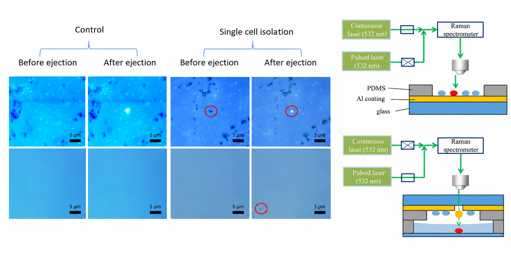
AI-assisted composite. Control and single-cell isolation views before and after ejection are shown alongside the Raman measurement and cell-ejection workflow. This AI-assisted composite combines user-supplied images with an adaptation of Song et al. (2017); details may have been redrawn and should not be interpreted as unaltered experimental data.
User-supplied images; adapted from Song et al. (2017)Source figureSource figure: CC BY 4.0View larger ↗
From measurement to isolation
Faster sorting with stimulated Raman imaging
Spontaneous Raman measurements provide rich chemical information, but signal collection can limit sorting speed. Stimulated Raman-activated cell ejection (S-RACE) combines faster chemical imaging, image analysis and laser-based ejection in one workflow. Cells are selected according to their Raman signatures and physically collected for further investigation.
The 2024 study separated lipid-rich yeast from a mixed population at approximately 13 cells per second, with the sorting result checked by downstream quantitative PCR. It also demonstrated recovery and cultivation of selected microorganisms and compatibility with tissue sections. Throughput, purity and recovery depend on the sample and operating conditions; the reported rate describes the tested yeast-sorting experiment.
Featured publications
- Raman microscopic analysis of single microbial cellsFoundational Report
Analytical Chemistry (2004)
- Raman-FISH: Combining stable-isotope Raman spectroscopy and fluorescence in situ hybridizationRaman-FISH
Environmental Microbiology (2007)
Environmental Microbiology (2018)
Microbial Biotechnology (2017)